This web page was created programmatically, to learn the article in its unique location you possibly can go to the hyperlink bellow:
https://www.westbrowardeyecare.com/retinal-photography-vs-dilation-modern-eye-exams-in-tamarac/
and if you wish to take away this text from our web site please contact us
When you visit your eye doctor for a complete examination, it’s possible you’ll encounter two distinct approaches for evaluating the again of your eye: conventional pupil dilation or superior retinal imaging know-how. As healthcare customers change into more and more knowledgeable about their choices, understanding these examination strategies has by no means been extra vital.
Both strategies serve the vital goal of detecting eye illnesses early—typically earlier than signs seem—however they provide totally different benefits relying in your particular person wants, way of life, and well being profile. This complete information examines the science, advantages, and sensible concerns of every method that can assist you make knowledgeable selections about your eye care.
Understanding Retinal Photography (Digital Retinal Imaging)
Retinal pictures represents a big development in eye care know-how, using refined digital cameras to seize high-resolution photos of your eye’s inside constructions. The most widely known system is ultra-widefield imaging know-how, resembling Optomap, which might doc as much as 82% of the retina in a single seize lasting lower than half a second.
The Technology Behind Retinal Imaging
Modern retinal cameras make use of a number of laser wavelengths to create detailed, layered photos of eye constructions:
- Green laser (532 nm): Penetrates the sensory retina and retinal pigment epithelium layers
- Red laser (633 nm): Examines deeper constructions from the RPE to the choroid
- Blue laser (488 nm): Enhances coloration imaging and allows fluorescence pictures
- Infrared laser (802 nm): Facilitates specialised angiography procedures
This multi-spectral method permits eye care professionals to look at totally different retinal layers concurrently, offering complete diagnostic info that rivals conventional examination strategies.
Clinical Advantages of Retinal Photography
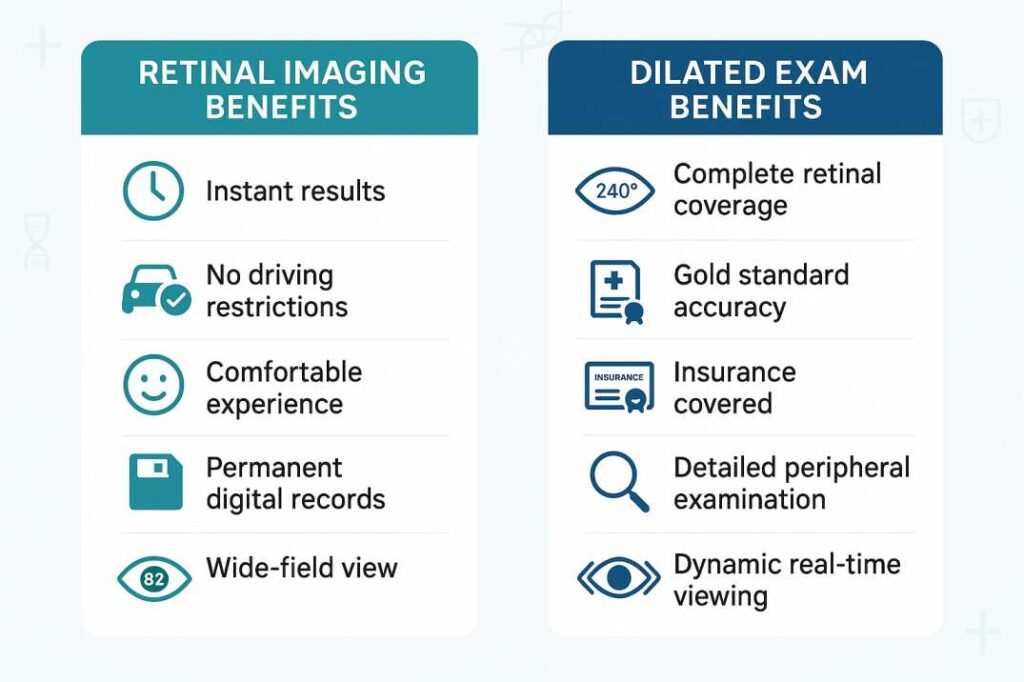
Immediate Documentation and Analysis: Digital photos can be found immediately for assessment, enabling real-time dialogue between physician and affected person. This quick availability enhances affected person training and permits for collaborative therapy planning throughout the identical go to.
Superior Patient Comfort The examination requires no eye drops, eliminates ready intervals, and produces no post-procedure imaginative and prescient results. Patients can drive instantly after the examination and resume regular actions with out restriction.
Comprehensive Field of View Advanced techniques seize as much as 97% of the retina utilizing auto-montage know-how, offering panoramic views that exceed the protection of many conventional examination strategies.
Long-term Monitoring Capabilities: Images change into a part of the everlasting medical document, enabling exact monitoring of retinal adjustments over time. This longitudinal documentation proves invaluable for monitoring illness development and therapy effectiveness.
Enhanced Diagnostic Accuracy Research demonstrates that retinal imaging can detect refined adjustments that may be missed throughout routine examinations, notably microaneurysms, small hemorrhages, and early drusen formation related to macular degeneration.
Comprehensive Analysis of Dilated Eye Examinations
Pupil dilation stays the foundational approach for complete retinal analysis, having served because the scientific normal for over 5 many years. This technique makes use of specialised medicines to briefly enlarge the pupils, offering eye care professionals with direct visualization of inside eye constructions.
The Dilation Process: Medical Precision
During a dilated examination, topical medicines resembling tropicamide or cyclopentolate are utilized to chill out the iris muscle tissues and briefly paralyze the attention’s focusing mechanism. This pharmacological intervention usually requires 15-Half-hour to realize full impact and might final 4-6 hours.
Unique Advantages of Dilation
Complete Retinal Coverage Dilation allows examination of as much as 240 levels of the retina, together with the far periphery the place retinal tears and detachments generally happen. This complete protection stays unmatched by present imaging know-how.
Dynamic, Real-time Examination. Unlike static imaging, dilation permits for dynamic examination the place the physician can regulate viewing angles, give attention to particular areas of concern, and carry out detailed stereoscopic analysis of retinal constructions.
Superior Optic Nerve Assessment The three-dimensional view supplied by means of dilated pupils gives optimum analysis of optic nerve well being, essential for glaucoma analysis and monitoring.
Essential for Pediatric Care. In kids, dilation stays vital for correct refractive measurements, because it briefly paralyzes the focusing muscle that may intervene with prescription willpower.
Insurance Coverage and Accessibility Dilated examinations are universally coated by medical health insurance plans as a part of normal eye care, making this feature accessible no matter monetary constraints.
Comparative Analysis: Making the Right Choice
Convenience and Lifestyle Factors
Retinal Imaging Advantages:
- No preparation or restoration time required
- Immediate return to regular actions
- Ideal for busy professionals and oldsters
- Suitable for sufferers delicate to medicines
- Comfortable for all ages, together with younger kids
Dilation Advantages:
- No extra value concerns
- Widely accessible in any respect eye care amenities
- Proven monitor document of scientific effectiveness
- No know-how dependencies or tools limitations
Diagnostic Capabilities Comparison
Detection Accuracy: Recent scientific research point out that combining retinal imaging with conventional examination strategies will increase lesion detection charges by roughly 30% in comparison with both technique alone. However, every method gives distinct diagnostic strengths:
Retinal Imaging Excels At:
- Early detection of diabetic retinopathy adjustments
- Monitoring macular degeneration development
- Documenting refined retinal hemorrhages
- Tracking therapy response over time
- Patient training and engagement
Dilation Remains Superior For:
Economic Considerations
Retinal Imaging Costs:
- Typical charge vary: $29-60 per examination
- Usually not coated by imaginative and prescient insurance coverage
- May be coated by medical insurance coverage when medically indicated
- Some practices embrace imaging in complete examination charges
Dilation Costs:
- Covered by most insurance coverage
- No extra affected person value
- Part of a typical complete eye examination
- Universal accessibility no matter monetary standing
Evidence-Based Research and Clinical Outcomes
Recent Scientific Developments
Study 1: Enhanced Diabetic Retinopathy Detection (2024) Research revealed in retinal imaging journals demonstrates that ultra-widefield imaging identifies 19% extra diabetic retinopathy instances in comparison with conventional seven-field pictures. When the total 200-degree view was utilized, 15% of instances acquired increased severity grades, doubtlessly resulting in earlier intervention and higher outcomes.
Study 2: Comprehensive Lesion Detection Analysis A landmark research from the New England College of Optometry revealed that combining retinal imaging with dilated examination elevated total retinal lesion detection by 30% in comparison with conventional ophthalmoscopy alone. Detection charges for particular pathologies improved dramatically:
- Drusen detection: 90-100% (image-assisted) vs. 15-62% (conventional alone)
- Small retinal hemorrhages: 95% (image-assisted) vs. 45% (conventional alone)
Study 3: Ultra-Widefield Coverage Validation Current analysis confirms that trendy retinal imaging techniques seize 82% of the retina in normal mode, with auto-montage capabilities extending protection to 97%. Importantly, research point out that 66% of retinal pathology happens past the attain of ordinary fundus cameras, supporting the worth of wide-field imaging know-how.
Clinical Guidelines and Professional Recommendations
When Retinal Imaging Is Preferred
Ideal Candidates:
- Routine screening in wholesome adults
- Patients requiring frequent monitoring (diabetes, hypertension)
- Individuals with dilation contraindications
- Busy professionals with time constraints
- Children who could not cooperate with drops
- Patients requiring detailed documentation for referrals
Clinical Scenarios:
- Annual diabetic retinopathy screening
- Macular degeneration monitoring
- Baseline documentation for brand new sufferers
- Insurance or authorized documentation necessities
When Dilation Remains Essential
Critical Situations:
- First complete eye examination
- Symptoms of retinal detachment (flashes, floaters, imaginative and prescient adjustments)
- Glaucoma analysis and administration
- Pediatric refractive assessments
- Complex retinal pathology investigation
- Emergency eye examinations
High-Risk Populations:
- Patients with a household historical past of retinal illness
- Individuals with diabetes or hypertension
- High myopia sufferers
- Previous retinal surgical procedure sufferers
- Those taking medicines that have an effect on the retina
Age-Specific Recommendations and Guidelines
Examination Frequency by Demographics
Adults Under 40 (Low Risk):
- Comprehensive examination each 2-3 years
- Retinal imaging is acceptable for routine screening
- Dilation really helpful for baseline institution
Adults 40-64 (Moderate Risk):
- Comprehensive examination each 1-2 years
- A mixed method is usually optimum
- Annual dilation for diabetes or hypertension
Adults 65+ (High Risk):
- Annual complete examinations
- Dilation strongly really helpful
- Retinal imaging is effective for monitoring development
Pediatric Considerations:
- Age-appropriate examination frequency
- Retinal imaging most popular for consolation
- Dilation is important for correct prescriptions
Practical Decision-Making Framework
Questions to Guide Your Choice
Assess Your Risk Profile:
- Do you will have diabetes, hypertension, or a household historical past of eye illness?
- Are you experiencing new signs (flashes, floaters, imaginative and prescient adjustments)?
- Is this your first complete eye examination?
- Do you will have a historical past of retinal issues?
Consider Your Lifestyle:
- Do you will have vital actions deliberate after your appointment?
- Are you comfy with short-term imaginative and prescient adjustments?
- Can somebody drive you dwelling if wanted?
- Are you delicate to eye drops or medicines?
Evaluate Your Preferences:
- Is value a big consider your determination?
- Do you favor probably the most complete examination potential?
- Would you profit from visible documentation of your eye well being?
- Are you curious about monitoring adjustments over time?
Professional Consultation Guidelines
Your eye care skilled will take into account a number of elements when recommending an examination method:
- Medical historical past and present well being standing
- Previous eye examination findings
- Current signs or considerations
- Family historical past of eye illness
- Age and threat issue evaluation
- Lifestyle and scheduling concerns
The Future of Retinal Examination
Emerging Technologies
The discipline of retinal imaging continues to evolve quickly, with a number of promising developments on the horizon:
Artificial Intelligence Integration: AI-powered evaluation techniques are being developed to mechanically detect and classify retinal abnormalities, doubtlessly enhancing diagnostic accuracy and decreasing interpretation time.
Enhanced Imaging Modalities New applied sciences combining a number of imaging strategies (OCT, autofluorescence, angiography) in a single system promise much more complete retinal analysis.
Portable and Telemedicine Applications Smartphone-based retinal cameras and telemedicine platforms are increasing entry to retinal screening, notably in underserved communities.
Clinical Practice Evolution
Modern eye care more and more emphasizes personalised approaches that mix one of the best elements of conventional and superior strategies. This evolution displays rising recognition that optimum affected person care requires versatile, individualized methods reasonably than one-size-fits-all approaches.
Special Considerations for Florida Residents
Environmental and Lifestyle Factors
Living in Florida presents distinctive concerns for eye well being that affect examination selections:
UV Exposure Concerns The intense year-round daylight in Florida will increase dangers for cataracts and macular degeneration. Both examination strategies can detect UV-related retinal injury, however retinal imaging supplies superior documentation of progressive adjustments.
Hurricane Preparedness: Severe climate occasions can disrupt medical information and prescription availability. Retinal imaging creates everlasting digital information that may be essential if bodily paperwork are misplaced throughout storms.
Active Outdoor Lifestyle Florida’s local weather encourages outside actions which will enhance eye damage dangers. Regular complete examinations—whether or not by means of imaging or dilation—assist guarantee early detection of trauma-related adjustments.
Aging Population Demographics: Florida’s vital retiree inhabitants requires specific consideration to age-related eye illnesses. Dilation stays the gold normal for detecting glaucoma and superior macular degeneration on this demographic.
Professional Recommendations and Best Practices
Integrated Approach to Modern Eye Care
Leading eye care professionals more and more advocate for individualized examination methods that leverage the strengths of each applied sciences. This built-in method acknowledges that optimum affected person care requires flexibility and scientific judgment reasonably than inflexible adherence to a single methodology.
For Routine Screening: Retinal imaging supplies wonderful baseline documentation and affected person engagement alternatives, notably for youthful, wholesome adults.
For Comprehensive Evaluation: Dilation stays important for full evaluation, particularly in sufferers with signs, threat elements, or complicated medical histories.
For Ongoing Monitoring: The mixture of each strategies typically supplies probably the most complete evaluation, notably for sufferers with recognized eye circumstances requiring cautious surveillance.
Quality Assurance and Clinical Standards
Regardless of the examination technique chosen, a number of elements guarantee optimum care high quality:
Professional Expertise The talent and expertise of the inspecting doctor stay probably the most vital elements in correct analysis and acceptable therapy suggestions.
Equipment Quality and Maintenance Modern retinal imaging gadgets require common calibration and upkeep to make sure diagnostic accuracy.
Comprehensive Assessment Eye examinations ought to at all times embrace a number of elements past retinal analysis, together with visible acuity, intraocular stress measurement, and anterior section evaluation.
Follow-up and Continuity: Establishing relationships with certified eye care professionals ensures acceptable follow-up and coordinated care when wanted.
Additional Resources and Clinical References
Professional Guidelines and Standards
1. American Academy of Ophthalmology – Comprehensive Adult Eye Evaluation Preferred Practice Pattern Guidelines. Published within the Ophthalmology journal, these evidence-based pointers set up skilled requirements for complete eye examinations, together with frequency suggestions primarily based on age, threat elements, and well being standing.
2. National Eye Institute – Dilated Eye Examination Guidelines Get a Dilated Eye Exam | National Eye Institute Federal well being company steerage offering official suggestions for dilated eye examinations, together with procedural info and illness detection capabilities.
3. Clinical Research – Image-Assisted Retinal Examination Comparison of Image-Assisted versus Traditional Fundus Examination Peer-reviewed analysis revealed in Eye and Brain demonstrating enhanced lesion detection when retinal imaging is mixed with conventional examination strategies.
Professional Medical Societies
- American Optometric Association: Clinical apply pointers for complete eye care
- American Academy of Ophthalmology: Professional requirements and persevering with training assets
- International Council of Ophthalmology: Global views on eye care greatest practices
These assets present evidence-based info supporting the scientific suggestions and pointers offered on this complete evaluation. Healthcare professionals and knowledgeable sufferers are inspired to seek the advice of these authoritative sources for extra technical particulars and present analysis developments.
Conclusion
The alternative between retinal pictures and dilated eye examinations represents a big determination in trendy eye care—one which displays the broader evolution towards personalised, technology-enhanced healthcare. Both approaches supply distinct benefits, and understanding these variations empowers sufferers to make knowledgeable selections aligned with their particular person wants, preferences, and circumstances.
Retinal imaging know-how has revolutionized eye care by offering comfy, quick, and extremely detailed documentation of retinal well being. Its capacity to interact sufferers by means of visible training and create everlasting information for longitudinal monitoring represents vital advances in preventive care.
Dilated examinations proceed to function the scientific gold normal for complete retinal analysis, providing unparalleled entry to peripheral retinal constructions and dynamic evaluation capabilities that stay unmatched by present know-how.
The way forward for eye care possible lies not in selecting between these approaches, however in thoughtfully combining them to create individualized examination methods that optimize each diagnostic accuracy and affected person expertise. As know-how continues to advance and scientific understanding deepens, the mixing of conventional experience with modern instruments guarantees even higher outcomes for sufferers in search of to protect their imaginative and prescient all through their lives.
For sufferers contemplating their choices: Engage in open dialogue with certified eye care professionals who can assess your particular person threat elements, way of life wants, and preferences to advocate probably the most acceptable examination method in your circumstances.
For healthcare suppliers: Stay present with evolving applied sciences and analysis whereas sustaining give attention to individualized affected person care that mixes scientific experience with acceptable use of accessible diagnostic instruments.
The final objective stays unchanged: early detection and efficient administration of eye illnesses to protect imaginative and prescient and improve high quality of life. Whether achieved by means of cutting-edge imaging know-how, time-tested scientific strategies, or considerate combos of each, this goal continues to information the evolution of contemporary eye care.
This article supplies academic info for healthcare customers and shouldn’t exchange skilled medical session. Individuals ought to seek the advice of certified eye care professionals for personalised examination suggestions and therapy selections.
This web page was created programmatically, to learn the article in its unique location you possibly can go to the hyperlink bellow:
https://www.westbrowardeyecare.com/retinal-photography-vs-dilation-modern-eye-exams-in-tamarac/
and if you wish to take away this text from our web site please contact us