This web page was created programmatically, to learn the article in its authentic location you’ll be able to go to the hyperlink bellow:
https://www.sciencealert.com/award-winning-images-reveal-our-smallest-realms-of-life-in-epic-detail
and if you wish to take away this text from our web site please contact us
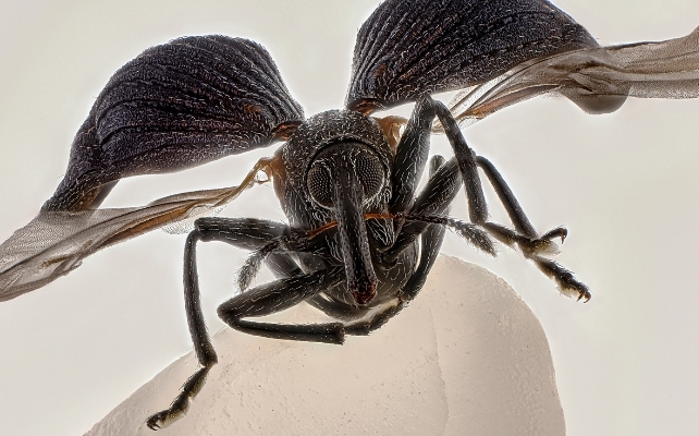
An astoundingly detailed weevil on a single grain of rice takes first place in 2025’s Nikon Small World photomicrography competitors.
Entomology fanatic Zhang You created this intricate picture by stacking over 100 fastidiously lit and cleaned photos collectively.
This approach permits the photographer to keep up a pointy focus throughout the depth of a topic, whereas a single picture will solely be targeted throughout a small vary of depth.
The complete course of took two weeks.
Related: Award-Winning Image Reveals a Hidden Culprit Behind Alzheimer’s
“It pays to dive deep into entomology: understanding insects’ behaviors and mastering lighting,” says You.
Insects play essential roles in our dwelling biosphere, from pollination to cleansing up natural waste.
While some bugs, like You’s weevil, are thought-about pests, many species at the moment are declining.
Such highly effective imaging can encourage us all to see the unbelievable complexity of their world and attempt to grasp them higher.
Now in its 51st 12 months, entries to Nikon’s extremely specialised pictures competitors proceed to awe us with the intricacies of existence on the microscopic scale.
Similarly exact and time-consuming efforts as You’s seemingly went into all 1,925 picture entries from 77 nations.
 Here are just a few others that caught our eyes.
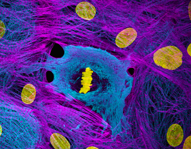
Tenth place went to a photograph of dividing coronary heart cells. When our cells put together to divide, they bundle up our genetic molecules (our chromosomes, proven in shiny yellow) earlier than mobile equipment pulls them into the 2 components of the fracturing cell.
We can see an instance of this bundling within the picture under, the place the chromosomes appear like a stack of bricks in the course of these grownup coronary heart cells, which have been grown from other adult cells.
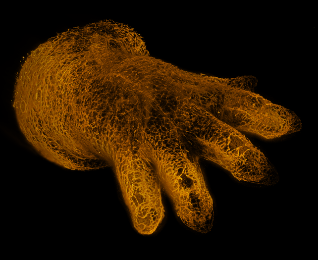
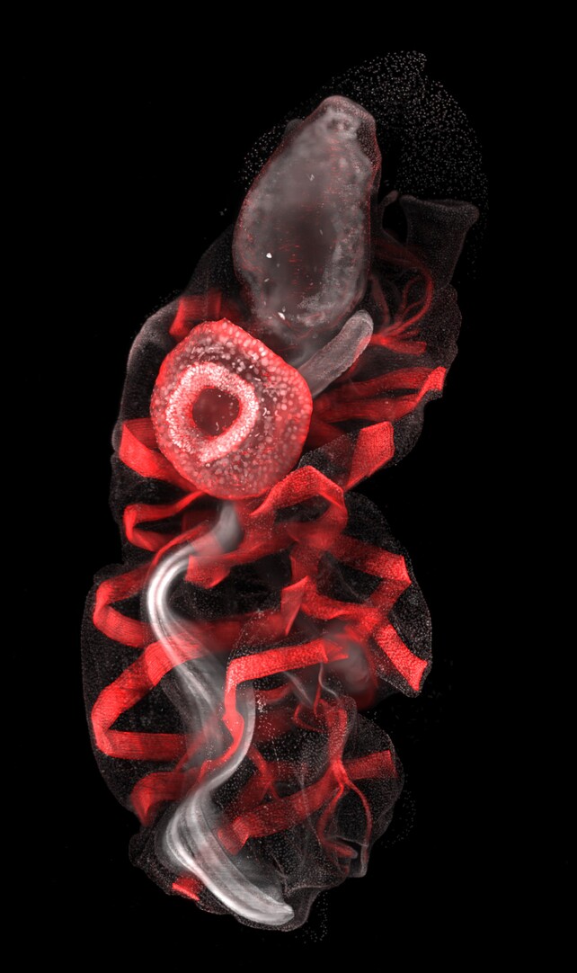
Disembodied vasculature within the form of a mammalian hand reaches out from the embryo of a growing mouse, on this eerie picture, which earned a distinction.
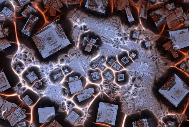
A disconcerting journey into the unusual alternate dimension of soy sauce fused with alum, a mixture of hydrated salts and metals typically used as a preservative, has us feeling dizzy.
This magnificent blob is an oozoid, the solitary asexual part of a sea squirt (Thalia democratica).
When it is able to reproduce, it can ooze out a protracted chain of clones, which grow to be females.
These females launch eggs that grow to be new oozoids, after which flip into males that launch sperm to fertilize different neighboring feminine chains.
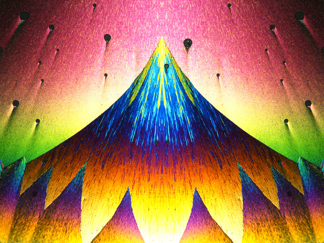
Polarized gentle brings out sudden shade combos and constructions on this picture of recrystallized phenyl imidazol, a substance typically used to assist create different chemical compounds.
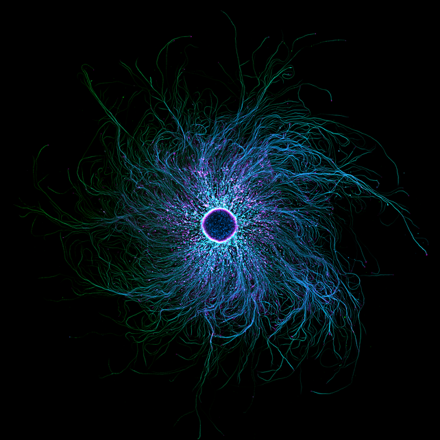
A swirling galaxy of fluorescently labeled mobile structural proteins, actin and tubulin, define the intricate branches of a sensory neuron. It’s other-worldly.
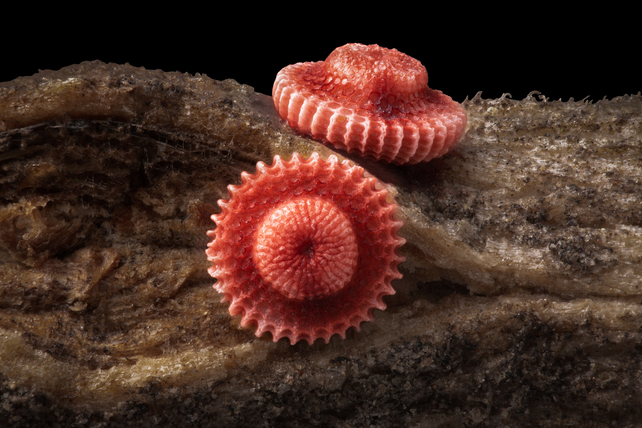
These delightfully odd pink latticed hats are the fragile little eggs of a butterfly, Artopoetes pryeri.
It’s a small white and black butterfly present in Asia, and the males have a dusting of blue to purple iridescence.
You can view many different entrants and winners here.
This web page was created programmatically, to learn the article in its authentic location you’ll be able to go to the hyperlink bellow:
https://www.sciencealert.com/award-winning-images-reveal-our-smallest-realms-of-life-in-epic-detail
and if you wish to take away this text from our web site please contact us