This web page was created programmatically, to learn the article in its unique location you possibly can go to the hyperlink bellow:
https://bmcmusculoskeletdisord.biomedcentral.com/articles/10.1186/s12891-025-08904-5
and if you wish to take away this text from our website please contact us
Study design
After acquiring institutional evaluation board approval, the research was performed from March 2024 to May 2024 at our hospital. A complete of 68 kids aged from 6 months to six years who have been suspected to have DDH underwent the radiographic examination. Written knowledgeable consent for participation and consent for publication have been obtained from all contributors’ guardians. All anteroposterior pelvic X-ray movies have been taken by a DR digital X-ray machine (DR Definium 6000 III, General Electric Company, United States). Participants with a historical past of hip surgical procedure, osteonecrosis of the femoral head, or extreme pelvic tilt have been excluded. Additionally, we employed the pelvic rotation index (PRI) and the pelvic tilt index (PTI) to evaluate the standard of pelvic X-rays [9, 10]. A radiograph with PRI and PTI values outdoors the conventional reference ranges was thought-about “non-standard” and was excluded from this research. Finally, we acquired 50 pelvic X-ray photos. The research flowchart is proven in Fig. 1.
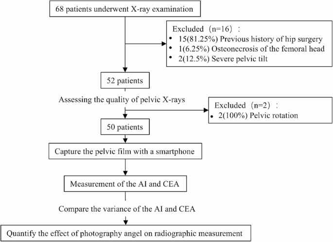
Flowchart of the research process
Photograph approaches
This experiment research primarily consisted of a movie viewer and a tripod. The movie viewer was positioned on the wall to make sure the movie was correctly plumbed, and the smartphone (Mi 14, Xiaomi Corporation, Beijing, China) was positioned horizontally on the tripod, which was mounted to the ground. Since the pictures have been taken indoors throughout the daytime, we turned off all gentle sources to keep away from reflections from the movie. In this experiment, pictures have been taken at completely different angles (tilt or rotation) utilizing an Android cellphone (Fig. 2). Tilting and rotation angles of the smartphone have been measured by a three-dimensional angle sensor (WT9011TCL-BT50, WitMotion, Shenzhen, China) with ± 0.2° static accuracy, linked by way of Bluetooth 5.0 at a 100 Hz sampling price. Tilt was outlined because the cellphone capturing up or down, the place up was (-) and down was (+). Smartphone`s rotation was achieved by the longitudinal axis of rotation of the tripod, the place left rotation was (-) and proper rotation was (+).
We positioned the pelvic movie on the movie viewer, opened the smartphone pictures utility software program, set it to regular pictures mode, turned on the reference line and crosshair capabilities, and used the utmost decision setting to take the image [5]. During the pictures course of, we aligned the crosshair of the picture`s display screen with the movie`s midpoint, which was decided and marked by a marker alongside the movie’s diagonal. Position info of the movie “R” was situated on the left facet of the pictures display screen. We set the pictures focal size to 50 mm and the capturing distance to 80 cm, as analysis signifies that underneath these situations, picture distortion is negligible [11]. Meanwhile, every of the 4 edges of the movie is saved parallel to the body, and the pictures taken underneath these situations are known as commonplace pictures.
After taking the usual pictures, whereas the cellphone is tilted, the highest and backside edges of the movie are saved parallel to the highest and backside frames of the picture. At the identical time, the tripod must be adjusted to make sure that the movie stays centered within the heart of the body. Likewise, throughout the rotation, we maintain the left and proper edges of the movie parallel to the left and proper edges of the picture. If the rotation angle exceeds a sure restrict, the movie could also be outdoors the picture. In such circumstances, the tripod could also be adjusted left or proper to reposition it centrally inside the {photograph}’s body, to take care of the deflection angle of the picture stays unchanged. Moreover, markers are used to attract three parallel strains of about 50 cm in size with a marker on the bottom beneath the tripod legs, and the tripod is restricted to motion alongside these strains. All obtained photos have a decision of roughly 4096(:occasions:)3072 pixels.
To keep away from extreme capturing angles that would end in out-of-focus photos or movie reflections, we thought-about particular person photographic habits that will result in the rejection of undesirable images. Consequently, we established the lean and rotation angles of the cellphone inside a spread of −20° to twenty°, with a gradient variation of 5°. This allowed for the acquisition of 17 units of images for every radiograph (Fig. 3), culminating in a complete of 850 images. Two orthopedic specialists evaluated the standard of the pictures. Images that have been blurred, out of focus, or exhibited extreme reflections have been excluded and required retaking. The closing chosen photos have been saved in JPG format. The unique photos have been additionally transferred to a pc and arranged utilizing a messaging app.

Standard pictures steps. The pelvic X-ray movie is mounted on the movie viewer, the smartphone is mounted horizontally on the tripod, the picture is going through the middle of the movie, and the 4 edges of the movie are saved parallel to the picture. Tilt and rotate the cellphone by the motion of the tripod

Photographic efficiency as a variance of the smartphone. a-b exhibits the view when the cellphone is tilted. c-d exhibits the view when the cellphone is rotated
Picture measurement
This research primarily investigates the impact of pictures angle variation on the measurement of the acetabular index (AI) and center-edge angle (CEA) in pediatric hip joints. AI is outlined because the angle between the road connecting the apexes of the bilateral Y-shaped cartilage (known as Hilgenreiner`s line) and the road drawn to the outer fringe of the acetabulum. CEA is measured utilizing Wiberg’s methodology of the CEA [9], which represents the angle between the road connecting the midpoint of the femoral head and the outer fringe of the acetabulum relative to the Perkin`s line (Fig. 4). Two skilled pediatric orthopedic surgeons measured the AI and CEA for every picture, repeating the measurements two weeks later. This was performed to evaluate each inter-observer and intra-observer reliability. The common of the 2 surgeons’ measurements was taken because the reference worth. Previous research documented measurement errors of AI and CEA have been 6° and 4° [12, 13], respectively. In this research, Measurement accuracy was outlined because the measurement error inside these clinically established thresholds. Changes under these validated thresholds have been thought-about clinically acceptable.

Method of measuring AI and CEA
Statistical evaluation
Continuous variables conforming to the conventional distribution have been reported because the imply ± commonplace deviation. Meanwhile, non-normal knowledge have been described by the median and the primary and third quartiles (Q1, Q3). We in contrast the outcomes of measuring parameters of pictures from completely different angles with the usual place (with out performing tilt or rotation operations), calculated the distinction between the 2, and quantitatively analyzed the adjustments in AI and CEA at completely different capturing angles. One-way ANOVA and Dunnett’s T3 take a look at have been used to evaluate the impression of pictures angle adjustments on two parameters. Additionally, linear regression evaluation was used to find out the connection between pictures angle adjustments and these two hip joint parameters. An impartial samples t-test was carried out to check variations in hip parameter adjustments between DDH and regular people. All statistical calculations and knowledge evaluation have been carried out utilizing SPSS (model 27), with p < 0.05 thought-about statistically important.
This web page was created programmatically, to learn the article in its unique location you possibly can go to the hyperlink bellow:
https://bmcmusculoskeletdisord.biomedcentral.com/articles/10.1186/s12891-025-08904-5
and if you wish to take away this text from our website please contact us