This web page was created programmatically, to learn the article in its unique location you possibly can go to the hyperlink bellow:
https://phys.org/news/2025-08-zebrafish-embryos-reveal-key-mechanisms.html
and if you wish to take away this text from our web site please contact us
. DOI: 10.1016/j.devcel.2025.07.011")
The heartbeat is synonymous with life. It’s one of many first important features to start throughout growth and to finish at loss of life.
“The heart is one of the first organs to form and function during development,” explains Rashmi Priya, head of the Crick’s Organ Morphodynamics Lab. “As the embryo grows, the heart continues to develop, expanding from a simple tube-like structure to a complex 3D pump.”
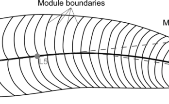
In new analysis revealed in Developmental Cell as we speak, scientists in Priya’s lab have examined a particular stage of this growth—the expansion of muscular ridges known as trabeculae.
As examine lead and postdoctoral fellow within the lab, Toby Andrews describes, “During early development, the inner surface of the heart forms a complex meshwork of these trabeculae. These structures must form in the right amount to ensure proper heart function. Defects in trabeculae formation are associated with several heart diseases called cardiomyopathies.”
Their work has revealed that the formation of trabeculae is pushed by suggestions between heartbeats and cell form adjustments.
A clear mannequin
To see the guts develop in actual time, the workforce turned to the Crick’s aquatic facility, residence to the zebrafish which are used to review early growth.
“Zebrafish hearts are very similar to ours in terms of structure and function, but their small size and transparency means we can use live imaging to watch developmental processes unfolding in real time as the heart beats inside the embryo—in this case, how the movements and shape changes of single cells build a functional organ,” explains Andrews.
Using highly effective microscopes and genetic instruments, they found that trabecular ridges develop not by cell division, as beforehand thought, however by transferring and rearranging the cells already in place.
They additionally discovered that the guts grows by stretching its present cells, virtually doubling in dimension, permitting the guts to broaden its quantity by 90% and maximize its blood filling capability.
Setting the proper tempo
Using computational fashions, the workforce uncovered a suggestions system the place the heartbeat and chemical alerts work collectively to regulate the density of those trabecular ridges, ensuring the guts develops in simply the proper strategy to perform correctly.
As the trabeculae develop and the guts contracts extra strongly, this initiates a mechanical sign that makes cells “softer,” enabling them to stretch and improve their dimension. This suggestions system dictates a wholesome tempo of development, as a result of as coronary heart cells stretch, they lose their means to get recruited, stabilizing trabecular development.
Priya describes this method as “self-organizing and intelligent,” the place the guts regulates its personal growth on the proper time, to fulfill the physique’s physiological calls for.
“The structure of the heart doesn’t stop changing at birth and continues to remodel over a lifetime,” provides Andrews. “Our hearts adapt their form and function to different physiological challenges, and a better understanding of the biology underpinning this flexibility could form the basis of future treatments for heart disease.”
Priya explains, “Even though we have come a long way in understanding the molecular pathways underlying cardiomyopathies, we still lack a clear picture of how trabeculae are shaped and how disruptions in trabecular architecture affect heart function. That’s why it’s crucial to uncover the core developmental programs that shape these ridges to enable the heart to function and support life.”
More info:
Toby G.R. Andrews et al, Mechanochemical coupling of cell form and organ perform optimizes coronary heart dimension and contractile effectivity in zebrafish, Developmental Cell (2025). DOI: 10.1016/j.devcel.2025.07.011
Citation:
Zebrafish embryos reveal key mechanisms behind lifelong coronary heart rhythm growth (2025, August 6)
retrieved 7 August 2025
from
This doc is topic to copyright. Apart from any truthful dealing for the aim of personal examine or analysis, no
half could also be reproduced with out the written permission. The content material is supplied for info functions solely.
This web page was created programmatically, to learn the article in its unique location you possibly can go to the hyperlink bellow:
https://phys.org/news/2025-08-zebrafish-embryos-reveal-key-mechanisms.html
and if you wish to take away this text from our web site please contact us