This web page was created programmatically, to learn the article in its authentic location you possibly can go to the hyperlink bellow:
https://www.salk.edu/news-release/how-can-scientists-visualize-cellular-life-with-greater-precision/
and if you wish to take away this text from our web site please contact us
How can scientists visualize mobile life with better precision?
- Highlights
- Salk researchers collaborated with scientists at Albert Einstein College of Medicine to develop a brand new class of probes for imaging residing cells
- The probes, referred to as visible-spectrum antigen-stabilizable fluorescent nanobodies (VIS-Fbs), generate high-contrast pictures with minimal disruption to regular mobile exercise
- The expertise allows extra exact investigation of advanced organic processes, together with cell signaling, improvement, and illness development
LA JOLLA—Fluorescent proteins have revolutionized science, enabling researchers to tag and visualize particular person molecules in residing cells, tissues, and animals. Using these instruments, researchers have watched viruses infect cells in actual time, noticed mobile trash assortment, and tracked the signaling that spurs tumor progress.
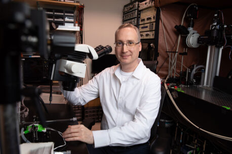
Click right here for a high-resolution picture.
Credit: Salk Institute
Salk scientists and collaborators at Albert Einstein College of Medicine have superior this visualization expertise. The new expertise, referred to as visible-spectrum antigen-stabilizable fluorescent nanobodies (VIS-Fbs), was validated in a number of mammalian cell varieties and supplies a strong instrument for a variety of life science analysis functions.
The examine was revealed in Nature Methods on April 22, 2026.
“This work establishes a versatile platform for imaging proteins with high specificity and minimal background,” says co-corresponding creator Axel Nimmerjahn, PhD, professor and Françoise Gilot-Salk Chair at Salk. “It opens new opportunities to study how molecular and cellular processes unfold in real time across diverse biological systems.”
How can present mobile imaging expertise be optimized?
The innovation started with tiny protein fragments referred to as nanobodies, which could be engineered to bind particular protein targets in residing cells. When fused to fluorescent proteins, these nanobody-based probes can reveal the place goal proteins are positioned and the way they behave. However, standard variations can nonetheless generate sign even when unbound, creating background fluorescence that may obscure fantastic particulars.

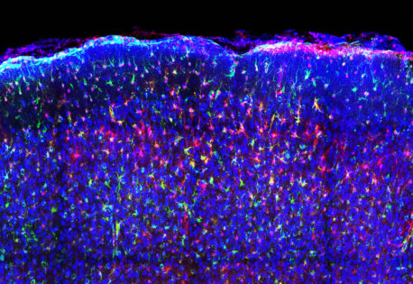
Click right here for a high-resolution picture.
Credit: Barykina et al., Nature Methods
The Salk and Einstein group designed a brand new kind of probe that retains the focusing on energy of nanobodies whereas vastly decreasing background fluorescence. VIS-Fbs change into steady and fluorescent solely when certain to their meant goal. This binding-dependent (“on-demand”) fluorescence reduces background noise by as much as a couple of hundredfold, enabling a lot sharper visualization of protein location and dynamics.
In addition, the researchers developed a number of variations of this new probe that fluoresce throughout almost the whole seen spectrum, from blue to far crimson. With this many coloration choices, a number of mobile targets could be tracked concurrently. Certain VIS-Fb variants will also be switched “on” and “off” with mild, making it potential to observe protein habits over time with excessive spatial and temporal precision. The researchers additionally established a modular design framework, enabling fast adaptation of VIS-Fb probes to completely different targets and purposeful readouts.
What do scientists examine with mild probes?
Click right here for a high-resolution picture.
Credit: Barykina et al., Nature Methods
The new expertise will permit scientists to realize extra correct, well timed perception into mobile exercise—even in advanced environments like residing mind tissue. The researchers demonstrated VIS-Fbs’ capabilities throughout a variety of residing fashions.
In mouse fashions, VIS-Fb probes enabled selective labeling and ratiometric imaging of calcium exercise in neurons and astrocytes throughout habits. In zebrafish, the expertise allowed real-time monitoring of dynamic modifications throughout early improvement and in response to medication that alter signaling pathways.
“Our results show that this imaging platform offers a much clearer and more precise view of how proteins behave inside living systems,” says co-corresponding creator of the examine Vladislav Verkhusha, PhD, professor and co-director of the Gruss Lipper Biophotonics Center at Albert Einstein College of Medicine. “It opens the door to studying complex biological processes, such as cell signaling, development, and disease progression, in new ways.”
Other authors and funding
Other authors embody Erin Carey of Salk; Natalia Barykina, Juliana Mendonça-Gomes, and Sofia de Oliveira of the Albert Einstein College of Medicine; and Olena Oliinyk of the University of Helsinki.
This examine was funded by the National Institutes of Health (GM122567, NS123719, GM147416), Jane and Aatos Erkko Foundation, Research Council of Finland, Finland Cancer Foundation, Chan Zuckerberg Initiative Foundation, NOMIS Foundation (Salk’s Neuroimmunology Initiative), and Edwards-Yeckel Research Foundation.
DOI: 10.1038/s41592-026-03056-3
This web page was created programmatically, to learn the article in its authentic location you possibly can go to the hyperlink bellow:
https://www.salk.edu/news-release/how-can-scientists-visualize-cellular-life-with-greater-precision/
and if you wish to take away this text from our web site please contact us