This web page was created programmatically, to learn the article in its unique location you possibly can go to the hyperlink bellow:
https://news.ufl.edu/2026/06/alzheimer-retina/
and if you wish to take away this text from our website please contact us
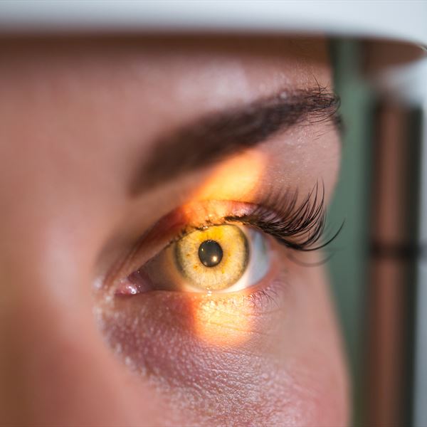
Often referred to as “the window to the soul,” the eyes may provide clues about one thing much less poetic however simply as necessary: the well being of the mind.
A brand new research of tens of hundreds of sufferers revealed that low-cost, easy and customary images of the retina behind the attention can precisely predict lots of the most typical danger components which can be related to growing Alzheimer’s illness.
“We know that Alzheimer’s disease develops over decades, but most of the diagnostic tools focus on late stage pathology when it is too late to intervene,” mentioned Ruogu Fang, Ph.D., a professor of biomedical engineering on the University of Florida who led the brand new research. “By looking at novel biomarkers, like retinal health, we offer new opportunities to identify patients at risk, offer appropriate tests and encourage them to develop healthy lifestyles to mitigate their risk.”
Fang and her collaborators, together with UF’s Adam Woods, Ph.D., and Meta researcher Yunchao Yang, Ph.D., published their findings June 16 within the Journal of Alzheimer’s Disease. The work was supported partially by the National Science Foundation.
Many sufferers routinely have footage of their eyes taken. Those with diabetes, glaucoma or cataracts can have many retinal images taken through the years. Even common eye exams for prescription glasses can seize photographs. That near-ubiquity makes analyzing retinal images easy and low-cost in comparison with different, dearer applied sciences like MRIs.
By utilizing machine studying to investigate these retinal images from greater than 40,000 sufferers in a United Kingdom-based affected person databank, Fang’s group was capable of establish areas of the retina related to Alzheimer’s danger components, such because the arteries and optical nerve.
“With the assistance of AI, we are now able to identify subtle retinal variations that were formerly overlooked across thousands of subjects, which may function as reliable indicators of future disease risk,” mentioned Seowung Leem, a doctoral scholar at UF and first writer of the publication.
The AI mannequin precisely predicted organic traits like intercourse or blood strain in addition to life-style components related to growing Alzheimer’s, similar to smoking, alcohol use and even insomnia. While many of those components are captured in sufferers’ medical charts, these data are sometimes incomplete. Some, like alcohol and smoking, depend on unreliable self-reports.
So retinal images might present one other, extra goal approach to detect these danger components. Plus, the retinal pictures can seize harm collected through the years, which can range between sufferers who share comparable danger components.
“Retinal morphology could provide measurable indicators of neurovascular integrity, which is highly relevant to Alzheimer’s disease vulnerability,” mentioned Fang, who can be affiliated with the McKnight Brain Institute. “In this sense, retinal imaging functions less as a surrogate questionnaire and more as an integrated biological sensor of cumulative risk.”
Fang’s group has already established that retinal photographs can detect active cases of Alzheimer’s disease. But scientists now consider the illness progresses over a few years, even many years. So figuring out early danger components may higher establish sufferers who may reply to earlier interventions — together with protecting life-style adjustments, some drugs and even mind coaching — earlier than irreversible harm to the mind takes place.
This web page was created programmatically, to learn the article in its unique location you possibly can go to the hyperlink bellow:
https://news.ufl.edu/2026/06/alzheimer-retina/
and if you wish to take away this text from our website please contact us